
CURRENT SLICE
Understands
the current slice
AI receives the open study context: current slice, viewing program, selected labels, zoom, and image position. Ask “What is marked here?” and get an answer for that exact frame.
Ask about anatomy, pathology, and the current slice. VIEWMIUM AI answers in atlas context and takes you back to the source image.
What is marked on the current slice?
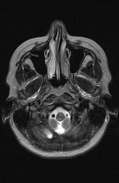 39404142CURRENT SLICE434445
VIEWMIUM AI receives the open study context—the current slice, selected labels, viewing program, and zoom—and connects its answer to atlas data.
Three anatomical structures are marked on the current slice:
Each link opens the related card or returns you to the relevant slice.
CURRENT SLICE
AI receives the open study context: current slice, viewing program, selected labels, zoom, and image position. Ask “What is marked here?” and get an answer for that exact frame.
ATLAS GROUNDING
Answers connect to published VIEWMIUM material. When atlas data is used, AI shows its sources instead of presenting a guess as fact.
DEEP LINKS
Links in an answer open the relevant module, study, structure or pathology card, and exact slice without making you search again.
ATLAS OCCURRENCES
Ask where a structure appears or how often it is annotated. AI gathers matches across published studies, groups slices, and shows a marked reference gallery.
VISUAL SEARCH
Upload a medical image, select the relevant region, and find visually similar published atlas frames. Open any result directly in the viewer.
EDUCATIONAL SAFETY
The assistant covers anatomy, radiologic anatomy, and educational pathology. It does not diagnose an image, prescribe treatment, or replace verification of clinically significant information.
Open a study, choose a slice, and ask a question—AI will keep the image context attached.
Launch the AI assistant→AI supports learning and does not replace clinical judgment.