
Step 01
Study submission
A clinician uploads a de-identified study or medical image series and adds its educational context.
Explore reviewed normal and pathology series with clickable annotations—or submit a de-identified study of your own for expert review and publication.
An educational platform for image-based learning and expert contribution.
One atlas, one continuous context
Catalog, full viewer, linked cards, comparison, and AI share one context. Switch modes to explore how the real product works.
 VIEWMIUMSearch the atlas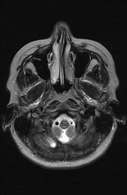
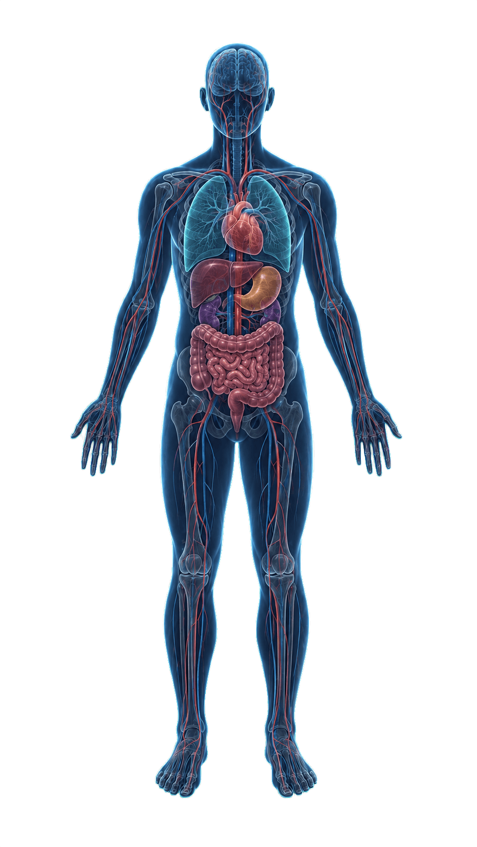
Expert-reviewed atlas growth
Clinicians can contribute educational normal and pathology studies. The team reviews each submission, helps prepare it, and adds it to the public atlas only after approval.
A clinician uploads a de-identified study or medical image series and adds its educational context.
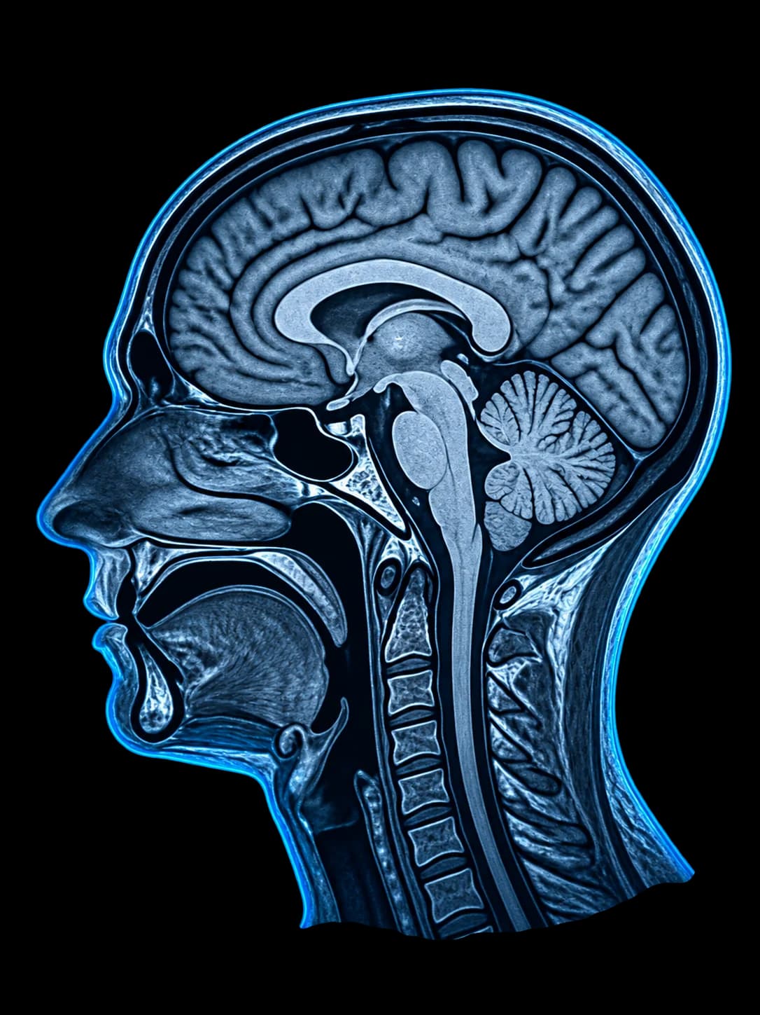
The case receives the description, key findings, and annotations needed for teaching.
A reviewer checks image quality, annotation accuracy, and publication safety.
An approved study becomes a complete interactive example for VIEWMIUM learners.
Who it’s for
Learn from reviewed material, use it for teaching, or help expand the atlas with educational studies of your own.
Who it’s for
Review normal anatomy and annotated findings inside complete studies of different types.
Open VIEWMIUMFAQ
VIEWMIUM is an educational platform with two connected parts: an interactive atlas of normal imaging and pathology, and a system where clinicians submit studies for review and publication.
The catalog contains reviewed normal and pathology series organized by anatomical region and modality. Annotations, cards, and educational notes vary by study.
Annotations are attached to a specific slice and area of the image. Tap one to open the related structure or pathology card.
A clinician uploads a de-identified study and adds educational context. The material undergoes medical and editorial review, and can enter the normal or pathology atlas only after approval.
No. VIEWMIUM is intended for education and reference. It does not provide medical advice and is not a diagnostic tool.
Keep exploring
Explore the atlas, submit a study, or understand how the platform works.
Upload a de-identified normal or pathology study and send it through expert review.
Submit a studyFollow the complete path from choosing a region to opening a linked anatomy or pathology card.
View the product tourLearn what is available in the atlas, how labels work, and which devices are supported.
Read common answers